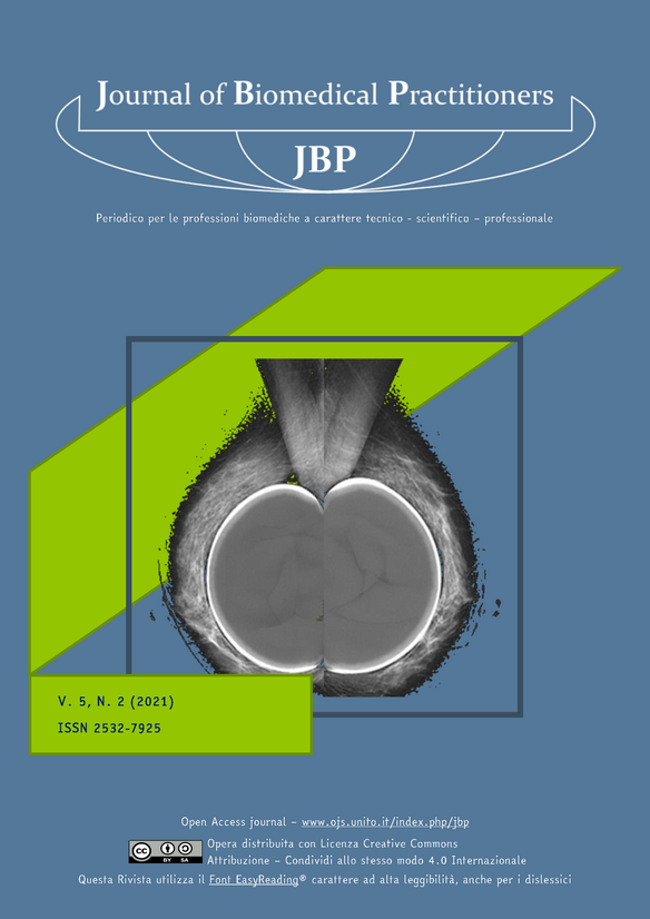
Study of the breast with implants in tomosynthesis
Main Article Content
Abstract
OBJECTIVE
Optimization of a low-dose protocol with Tomosynthesis technique for the study of breast with prosthesis during the screening process.
INTRODUCTION TO THE STUDY
Guidelines in both Italy, UK and US agree that 2D mammography, in the presence of prostheses, does not offer a reliable diagnosis and therefore invite clinicians to perform the Eklund maneuver. Due to the typical radiopacity of implants, Tomosynthesis is not currently the technique of choice for screening this type of breast. In this study, we wanted to experiment a protocol in which Tomosynthesis can be used to provide more diagnostic information even in the presence of breast implants, reconciling the technical, methodological and dosimetric aspects.
MATERIALS AND METHODS
A total of 53 women who joined the breast screening program reserved for women with prostheses aged between 50 and 74 years, in the ASL Roma 1, were enrolled in this study. These women were asked for consent to acquire the images also with Tomosynthesis.
In a first group of 15 women, using a standard configuration for the automatic exposure control algorithm (AEC), present on the IMS Giotto Class mammography unit, eight projections were acquired: four with digital mammography with prosthesis included and four in Tomosynthesis with Eklund's maneuver.
A low-dose configuration of the AEC was activated in a second group of 38 women. The average age of the women who took part in the study was between 50 and 65. One woman returned to the screening program 10 years after the quadrantectomy.
RESULTS
From the analysis and comparison of the data acquired in the different study modes, it emerged that the study in Mammography mode, with only four projections with prosthesis included, with standard exposure meter, is associated with an average glandular dose of 7.5 mGy.
If the Eklund maneuver is also performed in Tomosynthesis mode, the woman is exposed to an average of 11 mGy in total. A low-dose configuration of the AEC, for the eight projections, also acquired in tomosynthesis, is associated with an average glandular dose equal to 9.2 mGy total. This dedicated configuration allows to lower the dose by 15%, while maintaining adequate image quality.
DISCUSSION
At the present time, Tomosynthesis for the study of breasts with prostheses is not considered a valid diagnostic test yet, mainly due to the encumbrance of the prosthesis. However, the results of this test seem to highlight the possibility to use it effectively.
CONCLUSIONS
The study methodology tested is based on the best practices and on the most innovative technologies specifically designed for mammography. This methodology was able to improve the accuracy of screening tests, guarantee a controlled glandular dose and an adequate iconographic result. By disseminating this methodology, we may be able to increase the qualitative level of performance in the identified target.
Downloads
Article Details
The authors agree to transfer the right of their publication to the Journal, simultaneously licensed under a Creative Commons License - Attribution that allows others to share the work indicating intellectual authorship and the first publication in this magazine.
References
[2] Lavigne E, Holowaty EJ, Pan SY, Villeneuve PJ, Jhonson KC, Ferguson DA, Morrison H, Brisson J.; Breast cancer detection and survaival among women with cosmetic breast implant: systematic review and meta-analysis of ob-servational studies. BMJ. 2013; 346.
[3] A. Ciarrapico, A. Laghi, C. Pistolese, Nuove tecnologie, appropriatezza e costo efficacia dei programmi di pre-venzione di qualità, Rivista Tendenze nuove, ISSN: 2239-237otto, Il Mulino, art. 3/20013, pp 219-326, Tendenze nuove, ISSN: 2239-237otto, 219-326.
[4] Ekpo EU, Alkhras M, Brennan P. Errors in Mammography cannot be solved through technology alone, Feb 26;19(2):291-301Hughes LL, W. M. (2009 Nov 10;27 (32): 5319-24). Local excision alone without irradiation for ductal carcinoma in situ of the breast: a trial of the Eastern Cooperative Oncology Group. J Clin Oncol.
[5] Majid, Paredes et al, Missed breast carcinoma: pitfalls and pearls. 26 (2): 216–225. Fisher B. Land S. Mamounas E, e. a. (s.d.). Prevention of invasive breast cancer in women with ductal carcinoma in situ: anupdate of the Na-tional Sirgical Adjuvant Breast and Bowel Project experience. Semi. Oncol 2001; 2otto:400-41otto.
[6] Gibson DJ, Davidson RA. Exposure creep in computed radiography: a longitudinal study. Acad Radiol.2012 Apr; 19(4):45otto-62., Indicazioni ad interim per un utilizzo razionale delle protezioni per infezione da SARS-COV-2 nelle attività sanitarie e sociosanitarie (assistenza a soggetti affetti da covid-19) nell’attuale scenario emergen-ziale SARS-COV-2, Rapporto ISS COVID-19 n. 2/2020 Rev.

