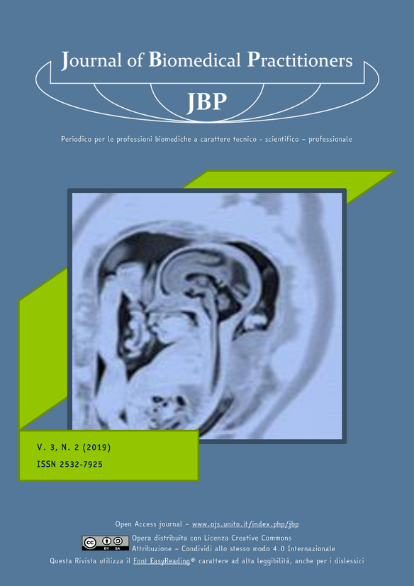
Risonanza magnetica fetale dell’encefalo: tecnica d'indagine e studio della correlazione tra età gestazionale e durata dell’esame
Contenuto principale dell'articolo
Abstract
Introduzione
In campo fetale la RM fu descritta per la prima volta nel 1980 per lo studio della placenta, ma solo negli anni ’90 si ipotizzò si potesse introdurre tale metodica per lo studio dell’encefalo fetale.
Gli ostacoli maggiori erano i tempi lunghi di acquisizione ed i movimenti fetali che creavano troppi artefatti. Per l’utilizzo elettivo nella pratica clinica si dovette aspettare l’implementazione delle sequenze “Single Shot” T2 - pesate ultraveloci.
Obiettivo dello studio
Verificare se esiste una relazione tra il tempo totale di esecuzione di un esame di Risonanza Magnetica Fetale (RMF) e l’età gestazionale della paziente al fine di poter ottimizzare la gestione dell’esame, diminuire ulteriormente i tempi di acquisizione ed aumentare la sicurezza della madre e del feto.
La RMF è una tecnica che non utilizza radiazioni ionizzanti, impiegata per lo studio del feto che, a partire dalle 19-20 settimane di gestazione, consente di ottenere importanti informazioni sull’anatomia e sullo sviluppo fetale ma deve essere considerata, oggi, un esame diagnostico di III livello che necessita di un quesito clinico mirato e giustificato posto dopo il risultato dell’indagine ecografica che è considerata di II livello.
Materiali e metodi
Lo studio è di tipo retrospettivo ed è stato condotto presso l’Unità Operativa di Radiologia e Neuroradiologia Pediatrica dell’Ospedale dei Bambini “Vittore Buzzi” di Milano.
Attraverso le funzioni statistiche del sistema RIS e PACS, sono stati raccolti e analizzati i tempi di esecuzione di un campione di 484 RM dell’encefalo fetale eseguite nel periodo tra gennaio 2015 e dicembre 2017.
Inoltre sono stati consultati i referti medici nel sistema RIS di tutte le RM al fine della raccolta del dato relativo all’età gestazionale della paziente.
Risultati
Dall’analisi dei dati si evince che, su 484 RM, il 30,37% è stato condotto in pazienti gravide alla 21° settimana di gestazione, l’8,68% alla 20° e l’8,26% alla 22° settimana; meno dell’1% ha interessato gravide sia alla 19° sia alla 35° settimana di gestazione.
Oltre la 31° settimana l’utilizzo di questa metodica è molto diminuito, in quanto non è più appropriata l’esecuzione della RMF ai fini della prognosi ed inoltre, spesso offre le stesse informazioni diagnostiche di una ecografia.
Sono stati calcolati ed analizzati i dati relativi a:- Tempo totale dell’esame successivamente suddiviso per età gestazionale
Dall’analisi dei dati si osserva come all’aumentare delle settimane di gestazione si riduca la durata dell’esame.
- “Tempo morto” (quello che intercorre tra le sequenze acquisite durante l’esame).
Dall’analisi dei dati emerge che esso tende a diminuire con l’avanzare dell’età gestazionale in quanto i feti più piccoli presentano maggiori movimenti per cui richiedono maggior abilità tecnica ed esperienza nell’impostazione dei piani di scansione delle sequenze.
- “Tempo vivo”, corrispondente al tempo di trasmissione delle radiofrequenze al feto.
Questo dato possiede un errore di calcolo intrinseco dovuto al fatto che è un tempo approssimato e varia a seconda delle dimensioni dell’encefalo fetale. Un feto più grande porterà ad un aumento del tempo vivo a causa del necessario incremento del numero delle “slice” di acquisizione.
- Numero totale delle sequenze acquisite.
Da questo dato si evince come la correlazione tra età gestazionale e numero di sequenze sia quasi inesistente. Tuttavia è doveroso considerare il limite di questa analisi, in quanto le sequenze che presentano in fase di acquisizione numerosi artefatti da non renderle diagnostiche, spesso non vengono archiviate nel PACS.
Conclusioni
Dai risultati ottenuti si può osservare come i tempi d’esame per lo studio dell’encefalo fetale abbiano la tendenza a diminuire con l’aumentare dell’età gestazionale.
Considerati i limiti dello studio, per una maggiore affidabilità dei risultati bisognerebbe analizzare ulteriori variabili non prese in considerazione nel presente studio in quanto emerse durante l’analisi dei dati, come ad esempio il quesito clinico o l’esperienza e competenza del TSRM, così da testare l’ipotesi che all’aumentare dell’esperienza pratica del TSRM possano ridursi i tempi di acquisizione dell’esame, in particolar modo per le gravide tra la 19° e 22° settimana di gestazione, che rappresenta la maggior sfida per la gestione dell’esame stesso.
Downloads
Dettagli dell'articolo
Gli autori mantengono i diritti sulla loro opera e cedono alla rivista il diritto di prima pubblicazione dell'opera, contemporaneamente licenziata sotto una Licenza Creative Commons - Attribuzione che permette ad altri di condividere l'opera indicando la paternità intellettuale e la prima pubblicazione su questa rivista.
Riferimenti bibliografici
Techniques, terminology, and indications for MRI in pregnancy, in “Seminars in Perinatology”; Bahado-Singh R.O, MD, Goncalves L.F,vol 37, 2013, pp. 334-339;
Prenatal magnetic resonance imaging: brain normal linear biometric values below 24 gestational weeks - C. Parazzini & A. Righini & M. Rustico & D. Consonni & F. Triulzi, Neuroradiology, 2008 Oct; (10): 877-83
Standard di Sicurezza in Risonanza Magnetica: Il Regolamento di Sicurezza; M. Giannelli & M. Mascalchi & M. Mattozzi & F. Campanella, Inail, versione aggiornata 2013
Indicazioni operative dell’Inail per la gestione della sicurezza e della qualità in Risonanza Magnetica
Temperature increase in the fetus due to radio frequency exposure during magnetic resonance scanning ; Iop Publishing - Physics In Medicine And Biology - Phys. Med. Biol. 2008 Nov 7; 53(21): L 15-8; P. A. Gowland and J. De Wilde
IEC 2008 Medical electrical equipment—part 2-33: particular requirements for the safety of magnetic reso-nance equipment for medical diagnosi
Safety of Mr imaging at 1.5 T in Fetuses: A Retrospective CaseControl Study of Birth Weights and the Effects of Acoustic Noise; B. Strizek et al; Radiology 2015 May; 275 (2): 530-7
Comparison Between 1.5-T and 3-T MRI for Fetal Imaging: Is There an Advantage to Imaging With a High-er Field Strength? Teresa Victoria Ann M. Johnson et al.- American Journal of Roentgenology 2016 Jan; 206(1): 195–201
Does 3 T fetal MRI improve image resolution of normal brain structure between 20and 24 week’s gestational age, Priego G. et al, American Journal of Neuroradiology August 2017, 38(8) 1636-1642
Dielectric effect artifact, Dr Matt A. Morgan et al, Radiopaedia
An ideal dielectric coat to avoid prosthesis RF- artefact in Magnetic Resonance Imaging, U. Zanovello et al, Sci Rep. 2017 Mar 23; 7(1): 326.
Elementi di risonanza magnetica: dal protone alle sequenze per le principali applicazioni diagnostiche; Co-riasco, Mario, Rampado, Osvaldo, Bradac, Gianni Boris (Eds.) – Springer, 2014
Manuale di RM per TSRM; Vanzulli, Torricelli, Cova, Cobelli, Colagrande, AAVV – Poletto Editore, 2013.

